CHECKLIST
FAVOURITES
BACKGROUND INFO
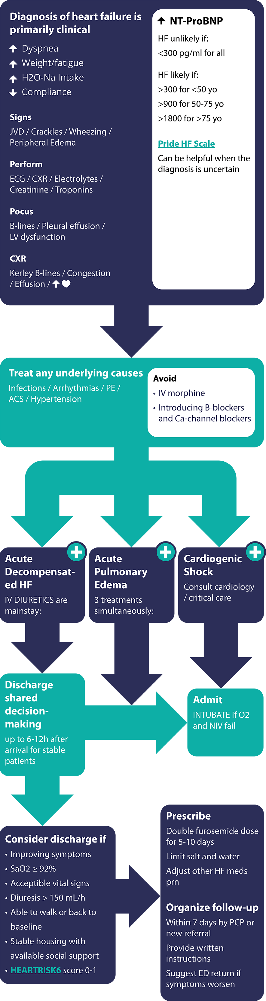
QUICK OVERVIEW

By using this app, you agree to these terms and conditions, which may from time to time be changed or supplemented by the Ottawa Hospital Research Institute or the Ottawa Hospital. If you do not agree to these terms and conditions, you should not use this app.
The medical information in this app is provided as an information resource only for health care professionals, to be used and applied at their own risk. This app and the information in it may supplement, but should not replace, the clinical judgement of a qualified health care professional. It is intended as a clinical decision-support tool only and is not a substitute for independent professional medical judgment.
This app and the information in it are not intended to be patient education, do not create any patient-physician relationship, and should not be used as a substitute for professional diagnosis and treatment. Please consult an appropriately qualified and licensed physician before making any healthcare decisions or for guidance about a specific medical condition.
The Ottawa Hospital Research Institute, The Ottawa Hospital and Dr. Ian Stiell expressly disclaim all warranties of any kind regarding the app and any information in it, either express or implied, including, without limitation, warranties of title, non-infringement, and implied warranties of merchantability or fitness for a particular purpose. The Ottawa Hospital Research Institute, The Ottawa Hospital and Dr. Ian Stiell shall have no liability for any damages, injury or any other loss whatsoever suffered as a result of your use of this app or the reliance on the information contained in it. In the case of loss, injury or damage of any kind to a third party resulting from your use of this app and the information in it, you agree to indemnify and hold harmless the Ottawa Hospital Research Institute, The Ottawa Hospital and Dr. Ian Stiell from any and all claims arising therefrom.
The content of this app is subject to copyright and trademark protection and should not be copied, reproduced, published or redistributed in any form without appropriate permission from the intellectual property owner.
1. What is acute heart failure (AHF)?
2. How does acute heart failure present?
3. Diagnosis of acute heart failure
1. What is acute heart failure (AHF)?
| Left ventricular ejection fraction (LVEF) in heart failure is classified as: | ||
| (HFpEF) | LVEF >50% |
| (HFmrEF) | LVEF 41%-49% |
| (HFrEF) | LVEF <40% |
| (HFimpEF) | compared to baseline |
PRIDE Acute Heart Failure Score4
| Predictor | Score | ||
| Interstitial edema on CXR | 2 | ||
| Orthopnea | 2 | ||
| Lack of fever | 2 | ||
| On loop diuretic | 1 | ||
| Age >75 years | 1 | ||
| Crackles/rales on lung exam | 1 | ||
| Lack of cough | 1 | ||
| Elevated NT-proBNP | 4 | ||
| |||
| Total | |||
| Likelihood of HF | Total Score |
| Low | 0-5 |
| Intermediate | 6-8 |
| High | 9-14 |
| NYHA Class | Definition |
| I | No symptoms |
| II | Symptoms with ordinary activity |
| III | Symptoms with less than ordinary activity |
| IV | Symptoms at rest or with minimal activity |
HEARTRISK6 Acute Heart Failure Risk Scale
| Points | |||
| 1. Initial Assessment | |||
| a.History of valvular heart disease1 | 1 | ||
| b.Heart Rate: | |||
| i. HR > 100bpm to <120 bpm | 2 | ||
| ii. HR >120 bpm | 3 | ||
| c.Treated with non-invasive ventilation2 | 2 | ||
| 2. Investigations | |||
| a.Creatinine: | |||
| i. >150 umol/L to <300 umol/L | 2 | ||
| ii. > 300 umol/L | 3 | ||
| b.Troponin: | |||
| i. > 3x or 4x upper limit of normal | 1 | ||
| ii. >5x upper limit of normal (initial or repeat, local hospital assay) | 2 | ||
| 3. Falls Reassessment after ED Treatment (2-6 hours) | |||
| a.Resting vital signs abnormal (SpO2 <90% on RA or usual O2, or HR > 110, or RR >28) |
1 | ||
| OR | |||
| b.Unable to start or complete 3-minute
walk test (vital signs become abnormal during walk)3 |
1 | ||
| TOTAL | |||
1moderate or severe valvular heart disease
2BiPAP within one hour of initial assessment
3score if patient SpO2 <90%, HR >110, RR >28 during walk test, or if unable
to complete due to fatigue or dyspnea
*no patient scored > 11
What is acute heart failure (AHF)? - Assessment
Acute Decompensated Heart Failure - Assessment
Acute Pulmonary Edema - Assessment
Isolated Right Ventricular (RV) Failure - Assessment
Cardiogenic Shock - Assessment
Etiology and Precipitating Factors - Assessment
History - Assessment
Clinical Exam - Assessment
Point-of-care Ultrasound (POCUS) - Assessment
ECG Indicators - Assessment
Investigations - Assessment
Chest X-ray - Assessment
The PRIDE HF Scale - Assessment
Overall Approach - Treatment
Acute Pulmonary Edema - Treatment
Isolated Right Ventricular Failure - Treatment
Cardiogenic Shock - Treatment
How to Decide if Patient Can be Discharged Home - Disposition and Follow-up
What is the HEARTRISK6 Scale? - Disposition and Follow-up
How to Optimize Discharge Medications - Disposition and Follow-up
What is Appropriate Follow-up? - Disposition and Follow-up
Heart failure risk categories for short-term serious outcomes
| Total Score | Absolute Risk | Category |
| 0 | 6.4% | Low |
| 1 | 8.5% | |
| 2 | 11.3% | Medium |
| 3 | 14.9% | |
| 4 | 19.4% | |
| 5 | 24.8% | |
| 6 | 31.2% | High |
| 7 | 38.3% | |
| 8 | 48% | |
| 9 | 53.9% | |
| >10* | 61.6% |
OXYGEN – NIV or high-flow cannula
IV DIURETICS – as in ADHF
OXYGEN consider NIV-intubation, safely sedate
Background and Methods
We created the CAEP Acute Heart Failure Best Practices Checklist to assist emergency physicians in Canada and elsewhere manage patients who present to the emergency department (ED) with acute heart failure. While there are several excellent cardiology society heart failure guidelines, there is nothing specific for ED management. The checklist attempts to fill that gap and focuses on patients presenting with one of four heart failure syndromes, acute decompensated heart failure, acute pulmonary edema, isolated right ventricular failure, and cardiogenic shock. There are detailed sections on diagnosis, treatment, disposition and follow-up.
The methodology and the format are similar to that of the 2021 CAEP Acute Atrial Fibrillation/Flutter Best Practices Checklist.11 We chose to adapt, for use by emergency physicians, existing high-quality clinical practice guidelines previously developed by the Canadian Cardiovascular Society (CCS).1 12 These guidelines were developed and revised using a rigorous process that is based on the GRADE (Grading of Recommendations Assessment, Development and Evaluation) system of evaluation.13 With the assistance of our PhD methodologist (IDG), we used the recently developed Canadian CAN-IMPLEMENT© process adapted from the ADAPTE Collaboration.14 15 We also reviewed heart failure guidelines from the European Society of Cardiology and the American College of Cardiology/American Heart Association/Heart Failure Society of America.2 9 We created an Advisory Committee consisting of 16 academic emergency physicians, three community emergency physicians, seven cardiologists, one general internist, three PhD methodologists, and two patient partners. The checklist was prepared and revised through a process of iterative feedback and discussions on all issues by all panel members. There were nine rounds of revisions until consensus was achieved. We then circulated the draft checklist for comment to approximately 500 Canadian emergency medicine and cardiology colleagues. Finally, the CAEP Standards Committee posted the Checklist online for all CAEP members to provide feedback. Only minor changes resulted from this feedback. The document has been approved by the CAEP Board.
While the panelists did offer the option of high dose nitrates for acute pulmonary edema, they chose not to use the term “SCAPE” (sympathetic crashing acute pulmonary edema) because it is not widely used and because the supporting evidence is weak.
Our hope is that the CAEP Acute Heart Failure Best Practices Checklist will standardize and improve care of acute heart failure in large and small EDs alike.
Advisory Committee Members
Academic Emergency Medicine | |
| Ian Stiell (Chair) | Ottawa Hospital, ON |
| Patrick Archambault | Hôtel-Dieu de Lévis, Lévis, QC |
| Bjug Borgundvaag | Sinai Health, Toronto, ON |
| Alexis Cournoyer | Hôpital du Sacré-Cœur, Montréal, QC |
| Kerstin de Wit | Kingston HSC, ON |
| Debra Eagles | Ottawa Hospital, ON |
| Andrew McRae | Foothills Hospital, Calgary, AB |
| Judy Morris | Hôpital du Sacré-Cœur, Montréal, QC |
| Robert Ohle | Health Sciences North, Sudbury, ON |
| Jeff Perry | Ottawa Hospital, ON |
| Brian H. Rowe | U of Alberta Hospital, Edmonton, AB |
| Frank Scheuermeyer | St Pauls Hospital, Vancouver, BC |
| Brian Steinhart | Unity Health, Toronto, ON |
| Alain Vadeboncoeur | Montreal Heart Institute, QC |
| Krishan Yadav | Ottawa Hospital, ON |
| Justin Yan | London HSC, ON |
Community Emergency Medicine | |
| Rupinder Sahsi | Grand River Hospital, Kitchener, ON |
| Troy Tebbenham | Peterborough Regional Health Center, ON |
| Suneel Upadhye | Niagara Falls, ON |
Cardiology/Internal Medicine | |
| Aws Almufleh | Kingston Health Science Centre, ON |
| Darshan Brahmbhatt | Sinai Health, Toronto, ON |
| Nadia Bouabdallaoui | Montreal Heart Institute, QC |
| Heather Clark | Ottawa Hospital, ON |
| Caroline McGuinty | Ottawa Heart Institute, ON |
| Robert J. Miller | Libin Cardiovascular Institute, Calgary, AB |
| Guillaume St-Pierre | Hôtel-Dieu de Lévis, Lévis, QC |
| Amelia Yip | St Mary’s Hospital, Kitchener, ON |
Methodology (PhD) | |
| Ian D. Graham | Ottawa Hospital Research Institute, ON |
| Stuart Nicholls | Ottawa Hospital Research Institute, ON |
| Sophie Boisvert | Université du Québec, Lévis, QC |
Patient Partners | |
| Marc Bains (Patient) | HeartLife Foundation |
| Christian Chabot (Patient) | Quebec City, QC |
References
Acknowledgements
Funding for this guideline was provided by The Ottawa Hospital Academic Medical Organization (TOHAMO).
No authors declare competing interests.
We thank the hundreds of Canadian emergency physicians and cardiologists who reviewed the draft guidelines and who provided very helpful feedback. We thank Ottawa Hospital Research Institute Emergency Research staff for their assistance: Angela Marcantonio, Catherine Clement, Carolyne Kennedy, Isabella Welch.